
Over the years, technology has significantly shaped the evolution of modern medicine. Advancements in devices, treatment methods, data processing, and diagnostic capabilities have paved the way for many of the innovations healthcare professionals rely on today.
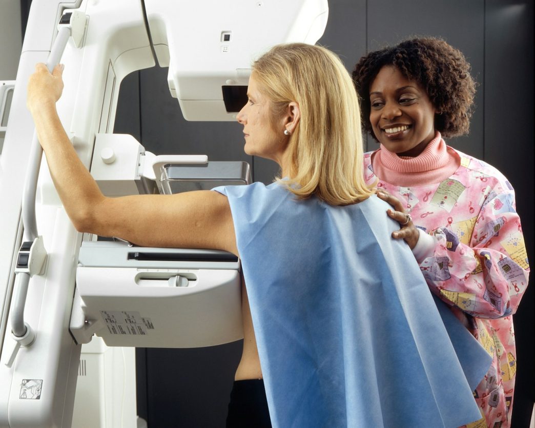
In the field of breast imaging, technology has been instrumental in making the change from analog film to advanced digital and artificial intelligence (AI) systems. Due to these advancements, the detection rate of invasive cancers has increased, and patient recall rates have dropped significantly in recent years.
While it’s easy to get used to today’s technology, we should also make sure to remember where we came from, especially those who hope to work in the field. So, let’s have a look behind the scenes and see how technology changed mammography.
The Evolution of Mammographies
Before digital sensors, mammograms were captured strictly on specialized photographic film. This meant that images could not be adjusted after exposure, so any mistakes meant a re-take, thus prolonging the diagnosis time. Plus, dense breast tissue and tumors both appeared white on film, making it difficult to identify any early-stage cancers.
In the early 2000s, with the help of solid-state detectors, technicians were able to convert X-rays into electrical signals, creating digital images. This allowed radiologists to adjust contrast, brightness, and magnification electronically, greatly reducing the rate of technical repeats.
The evolution of breast imaging continued during the following years, with the first Digital Breast Tomosynthesis (DBT) system, which captures multiple low-dose exposures. A computer then reconstructs these images into thin, 1-millimeter slices, eliminating the problem of overlapping tissue hiding a tumor or mimicking a false alarm.
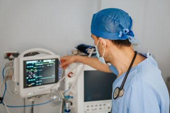
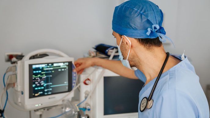
Nowadays, technology is intertwined with the medical field, improving diagnostics, allowing remote patients access to services and specialists, and making at-home healthcare a possibility. For breast imaging, advanced deep learning algorithms act as a second pair of eyes for radiologists, reducing the time to diagnostic and eliminating false positives.
How Digital Technology Changed Medical Careers
Digital technology has completely rewritten the job descriptions of all healthcare professionals. To become a mammographer, for example, you no longer need physical mechanics knowledge and darkroom chemistry. But you do need to be well-versed in advanced data management, computational analysis, artificial intelligence collaboration, and highly specialized patient care.
In the analog era, a mammography technologist had to know how to capture and interpret two flat views of each breast.
Today, the same technologist has to understand how to:
- Acquire optimal 3D data slices
- Use advanced software that generates a standard 2D image directly out of the 3D data
- Review the generated images for quality control
- Prep patients for and administer a Contrast-Enhanced Mammography (CEM)
- Interact with AI-driven Computer-Aided Detection (CAD) systems
- Identify and eliminate software errors that might trick the AI system into false positives
The Future is Promising
Whether it’s breast imaging or another branch of healthcare, the future is indeed promising. Current technology is already advanced enough to reduce data processing times and help with challenging diagnostics.
As AI-led systems improve, we might open the doors to more innovation and accelerate the development of safer, faster, and more effective healthcare solutions.